Stanford Says Your Brain Is Two Separate Organs. Is It?

Is your brain actually two separate organs? In September 2026 a Stanford study in Nature Neuroscience reported that the front of the brain (forebrain and midbrain) and the back (hindbrain) grow from two different groups of starter cells in the embryo, and the press release called the brain two organs. This video goes through what the experiments measured and where the interpretation begins: Nieuwkoop's 1952 model of a single neural sheet, cell tagging in mouse embryos, human stem cells that would not switch from front to back, the same split in a brainless acorn worm, the critics, comb jellies and jellyfish, the Hox-patterned hindbrain, lab-grown hindbrain motor neurons for ALS and SMA research, and why this is not the "lizard brain" story. It is about front and back, not left and right.
This article is the full narration of the video. Watch on YouTube
Is the brain two organs?
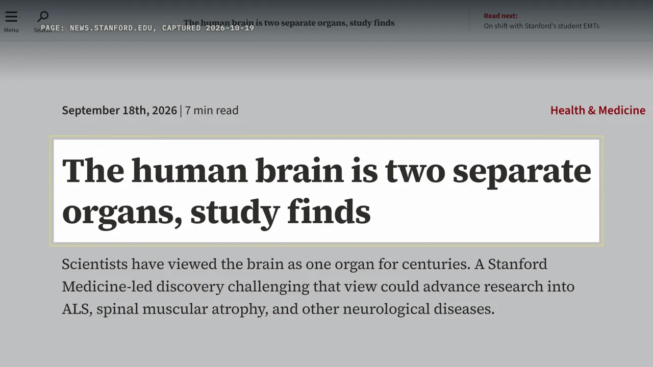
Is your brain actually two separate organs? In September 2026, that was the headline from Stanford University: the human brain is two separate organs, study finds. Smithsonian magazine put it more vividly: two distinct nervous systems that got squished together. Before anything else, this isn't about left brain and right brain, or split-brain patients.
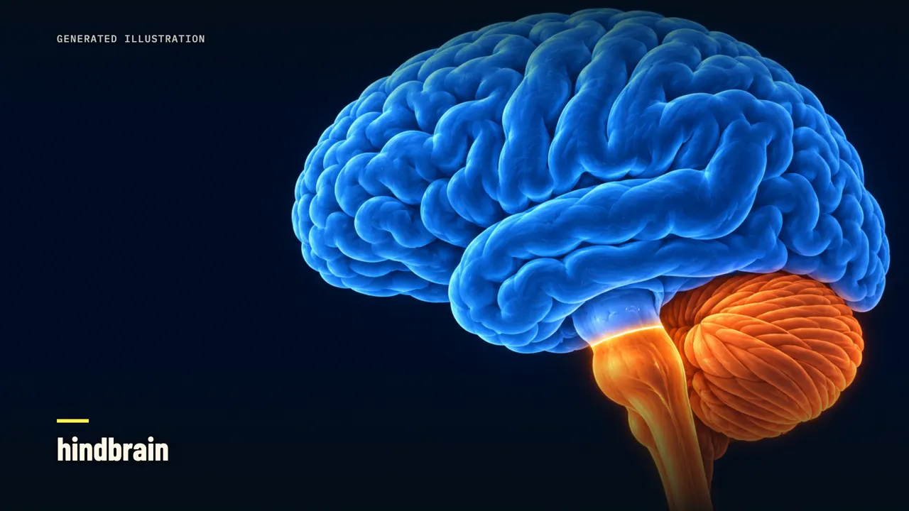
The split here runs front to back. On one side are the forebrain and the midbrain. On the other is the hindbrain, at the back and base of the skull, where the brain meets the spinal cord. The claim is that these two parts grow from two different kinds of starter cell.
Five days later, Ars Technica's science editor wrote: you do not have two brains. So who's right? Here's the short answer, before the long one. The experiments give strong evidence for two groups of starter cells, though they can't rule out a very brief common starter.
Calling those two organs is an interpretation, and even the lead scientist calls it only an idea. To see where the evidence ends and the idea begins, we have to start with a salamander experiment from 1952.
The textbook's single neural plate
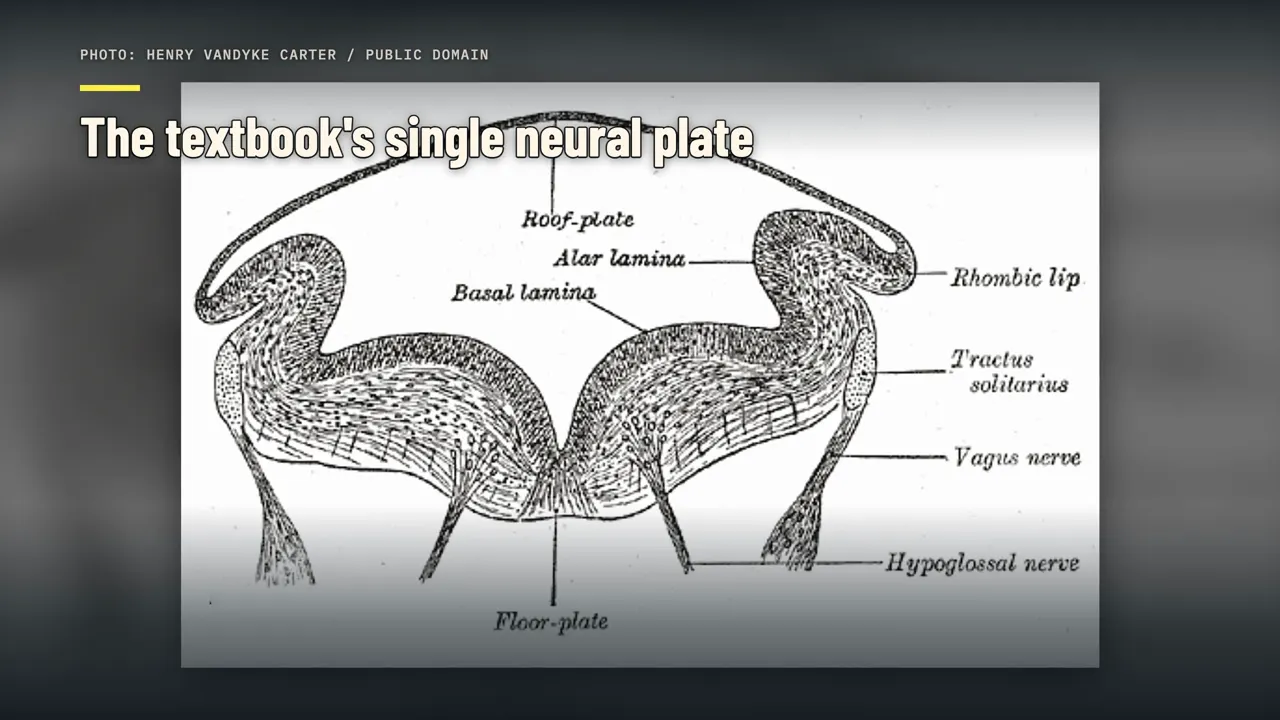
Every vertebrate brain starts as a flat sheet of cells on the back of the embryo, called the neural plate. That sheet rolls up into a tube, and the front end of the tube swells into the brain. So how does one sheet know which end becomes the forebrain and which becomes the hindbrain? In 1952, the Dutch embryologist Pieter Nieuwkoop tried to find out, using newt and salamander embryos.
He took flaps of outer tissue and pushed them into the embryo's neural tissue at different levels. The tip of each flap, farthest from the embryo, turned into forebrain tissue. Closer to the base, it turned into more posterior parts, matching the level where it was stuck in. From experiments like these, he proposed a two-step model.
In step one, a signal turns ordinary outer tissue into neural tissue. Left alone, that new neural tissue becomes only front structures, forebrain and midbrain. In step two, a second signal acts only on tissue that is already neural, and shifts it toward the back. The longer and stronger the exposure, the further back the tissue ends up: hindbrain, then spinal cord.
In this picture, there's one starting sheet, and the back of the brain is the front, transformed. A 1999 review said the model had survived to that day, and was still cited to explain new experiments. Nature's news story on the 2026 paper opens with the same picture. For decades, it says, many scientists have thought that a single type of starter cell gives rise to the entire brain.
Kyle Loh, the Stanford biologist who led the new work, compares it to the lung. It has two lobes, left and right, but nobody thinks it comes from two separate origins. And the brain's parts are joined end to end, so a single origin felt obvious. Two genes matter for the rest of this story: Otx2 and Gbx2.
In the mouse, Otx2 is on in the forebrain and midbrain, and Gbx2 in the front of the hindbrain. They meet at one border, between the midbrain and the hindbrain. In a 1999 study, mice without Gbx2 had the Otx2 zone spread backward. Switching Gbx2 on inside the Otx2 zone pushed that border forward, and the embryos grew a bigger hindbrain.
Loh says people had seen these two genes in different parts of the embryo back in the nineties. The new question goes earlier. Are the cells on each side of that border different from the very start?
Experiment one: tagging cells in a mouse embryo
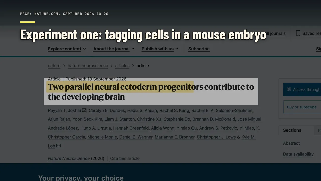
The study came from Loh's lab at Stanford and was published in Nature Neuroscience on September 18, 2026. By coincidence, Nieuwkoop died on September 18, 1996, exactly thirty years earlier. The full paper is behind a paywall, but an earlier version was posted openly in July 2025, so the numbers here come from that preprint. Where the published figures differ, I'll say so.
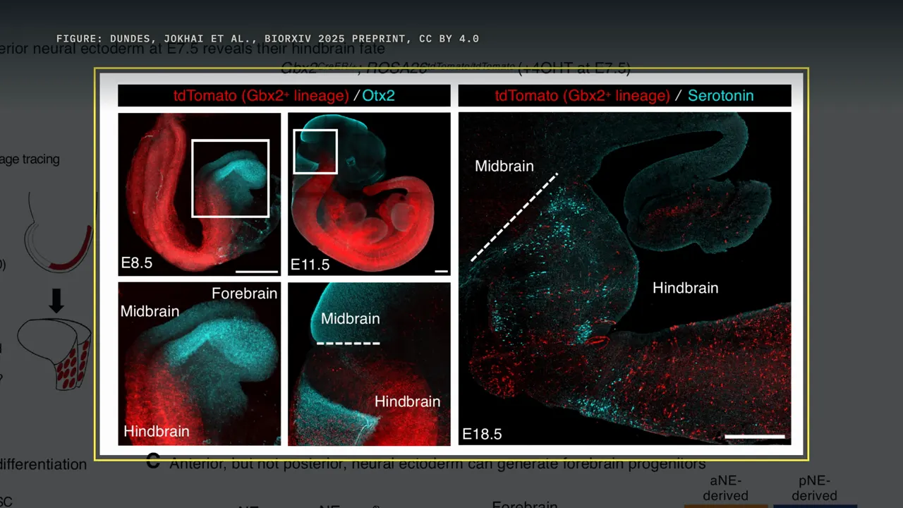
The first experiment followed cells over time in mouse embryos. The mice were engineered so that a single dose of a drug would permanently mark any cell that had Gbx2, the back gene, switched on at that moment. Each marked cell turned red, and so did every cell descended from it.
On day seven and a half of development, during gastrulation, the team gave that dose. Gastrulation is when the embryo lays down its basic layers, and it's when both kinds of starter cell first appear. Timing mattered here. An earlier study had tried something similar with tamoxifen, a drug that lingers in the body for several days.
That could tag hindbrain cells that only switched Gbx2 on later. This team used a short-lived version of the drug, so the tagging window lasted about twelve hours. One and two days later, the red cells were in the hindbrain, and none were in the forebrain or midbrain. Near birth, at day eighteen and a half, the red cells were still essentially absent from the forebrain and midbrain.
Marking half a day earlier, at day seven, gave the same pattern. But that test only follows cells that were already on the back side. So the team ran a second test that didn't pick a side. They used a genetic tool called Confetti, which marks single cells at random in one of three colors.
Each marked cell grows into a small cluster of the same color. Across 16 embryos, they counted 494 clusters that came from neural tissue. 311 of them, about 63 percent, were counted in the forebrain and midbrain. 161, about 33 percent, were counted in the hindbrain.
The authors describe each cluster as almost exclusively on one side or the other. That adds up to about 96 percent. The other 22 clusters, about four and a half percent, sat on both sides of the border. And the counting rule matters: a cluster with fewer than two cells across the border was counted on the side where most of its cells were.
If even one stray cell counted as mixed, the mixed share would be higher. Ars Technica made the obvious point: 96 percent is not 100 percent. Stanford's press release went further than the data, saying the two populations never overlap. Still, the authors' conclusion follows from the numbers: by the end of gastrulation,
most of these cells are already headed for the front brain or the back brain.
Experiment two: human stem cells that would not switch
Mice are not people, so the team also worked with human stem cells in a dish. They used several lines of human pluripotent stem cells, cells that can become almost any tissue. Within two days, using different mixes of signals, they turned them into either front-type or back-type starter cells. The back type formed when the team added FGF and a vitamin A-like retinoid, while blocking three other signals: BMP, TGF-beta and WNT.
Then came the key test. They gave back-type cells the signals that normally make forebrain and midbrain. Two days later, most of them hadn't switched. They gave front-type cells hindbrain signals, and those didn't switch either.
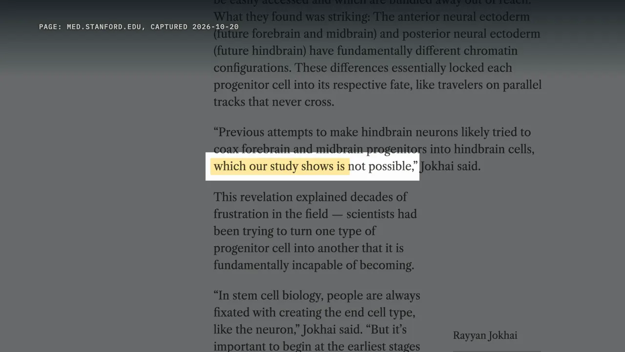
One of the two lead authors, a graduate student named Rayyan Jokhai, drew the practical lesson. Earlier attempts to make hindbrain neurons, he said, likely started from front-type cells, which the study shows is not possible. Still, the result wasn't absolute. When the two types were grown together, a few back-type cells switched on SIX3, a marker of the front of the brain.
Remember that gene; it comes back later. The team also looked at how each cell type packs its DNA, a layer of control called chromatin. Chromatin helps decide which genes a cell can easily use. The two cell types had clearly different patterns of open and closed DNA.
The authors read that difference as part of what holds each cell to its path. And they add their own caution: a dish may not recreate the full complexity of a real embryo.
Monkeys, chicks, fish and a brainless worm
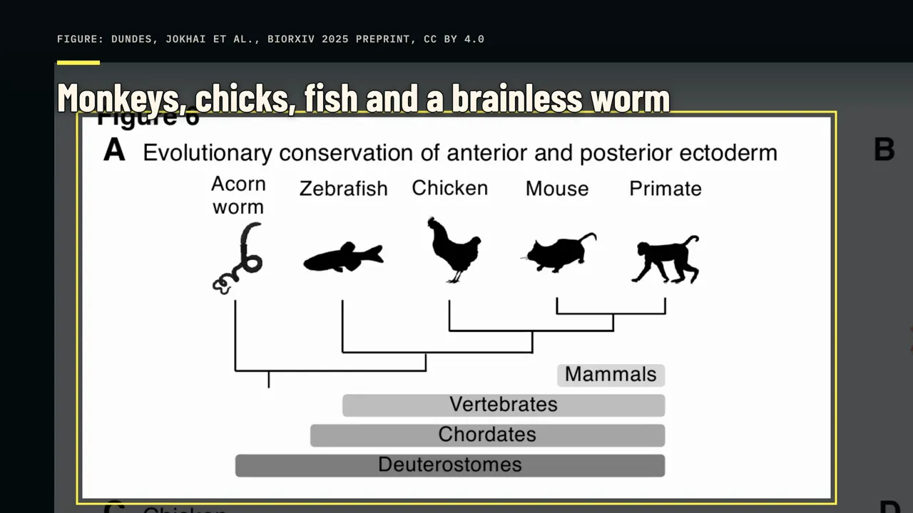
Next, the team asked whether this split is only a mouse and human thing. They reanalyzed published single-cell data from macaque, chicken and zebrafish embryos. In each one, they found the same two starter groups, front and back. Then they looked at an animal with no brain at all.
The acorn worm is a small burrowing animal of the seafloor, with only a scattered net of nerves. In 2003, a study found 22 genes that pattern the chordate nervous system switching on in its outer layer, in nearly the same front-to-back order as in animals with spinal cords. Christopher Lowe, an author of that study, is also on the 2026 paper.
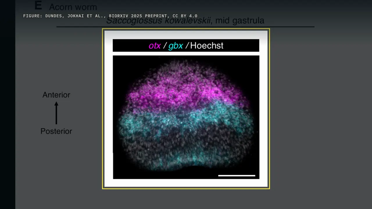
In gastrulating acorn worm embryos, the team saw the same two-part split. Here the published paper is stronger than the preprint. The preprint said those stains came from a single embryo per species. The published figures say the images represent five chicken embryos, nine zebrafish embryos and ten acorn worm embryos.
Acorn worms and humans last shared an ancestor more than half a billion years ago. The published abstract puts it at 550 million years, from hemichordates, the acorn worm's group, to mammals. Loh points out that's before the supercontinent Pangaea formed. If the split is that old, it would predate anything we'd call a brain in our lineage.
The pushback: is it really two?
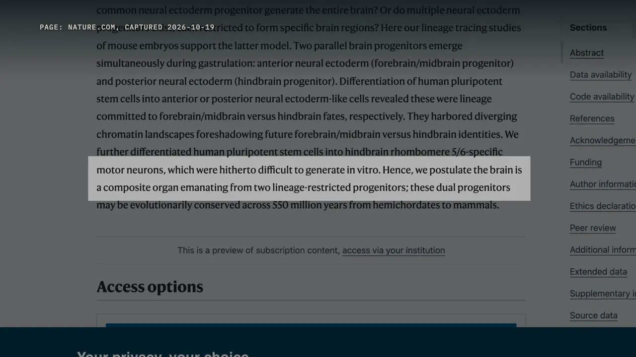
So does two starter groups mean two organs? The paper's abstract chooses its words carefully: we postulate the brain is a composite organ. Loh, in interviews, goes further. He told Nautilus that the idea of the brain as one organ is a historical anachronism.
His reasoning: scientists studied mice and humans first, where there's one skull with one big squishy gray thing inside. At the very least, he said, there are two separate organs. Stanford's release calls the brain two ancient nervous systems cleverly packaged together. But in October, Loh told Live Science that this is only an idea, and it might change as scientists learn more.
Outside scientists were more careful. Alex Pollen of UC San Francisco, who wasn't involved, raised one problem in Nature's news story. It's hard to prove, he said, that the two groups don't come from a single, short-lived starter cell. The authors agree that's possible.
They write that their results cannot exclude a brain-wide starter cell that exists for a very brief time. Cecilia Moens of the Fred Hutchinson Cancer Center isn't convinced the two fates are fully fixed. She has seen cells swap fates, even just for brief periods of time. Here too the authors leave the door open: whether the cells are truly committed inside a living embryo still has to be shown.
John Timmer at Ars Technica took a different angle. By the time the first cells committed to becoming neurons show up, he wrote, the embryo already knows its head from its tail, and its back from its belly. So it's not surprising that nerve cells inherit some sense of where they are.
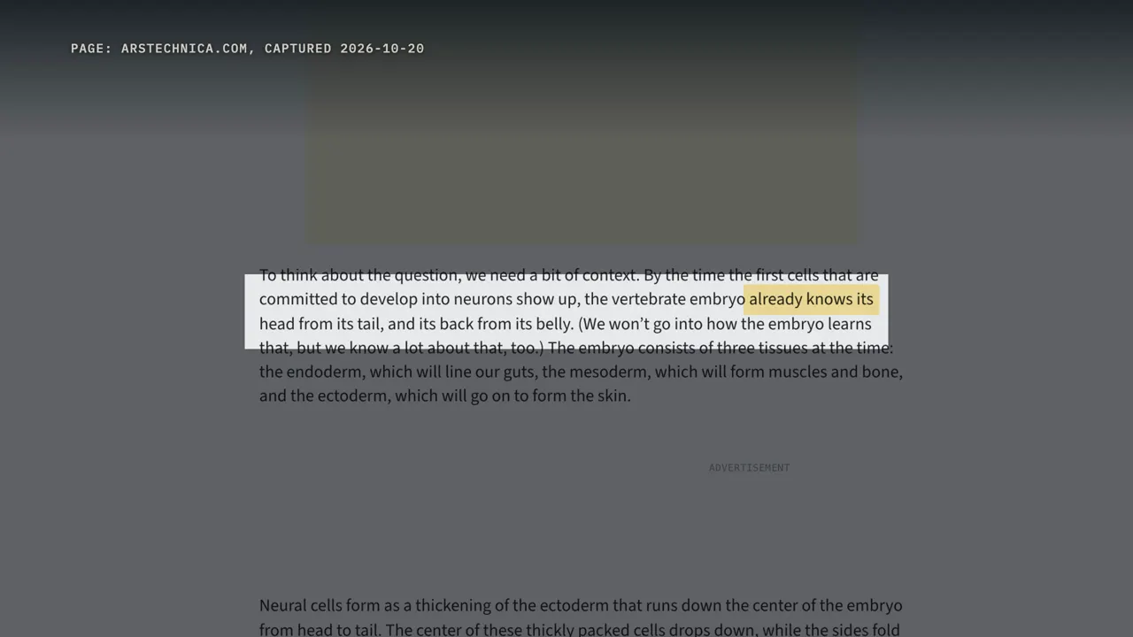
His verdict: you have one brain, with several distinct regions within it. There was harsher pushback, too. Loh says that years ago, after a talk he gave, the chair of a developmental biology department stood up and said: this is completely false. He called getting the paper published quite the uphill battle.
Others welcome it. Anna Di Cosmo of the University of Naples told Live Science it shows the deep ancestry of the vertebrate brain is fundamentally modular. So what does the word organ really mean here? The authors themselves point to the heart.
The heart grows from two cell groups, the first and second heart fields, which come together to form a single organ. Nobody says you have two hearts. And the spinal cord was already known to arise from separate starter cells of its own. So the paper's broader suggestion is that the whole central nervous system may be stitched together from several region-specific starters.
Loh says his lab is now testing the spinal cord directly. And on the cerebellum, he says there's simply no data either way. So the fair version is this: one brain, possibly built from two beginnings, and scientists are still arguing over what that means.
How many times did nervous systems evolve?
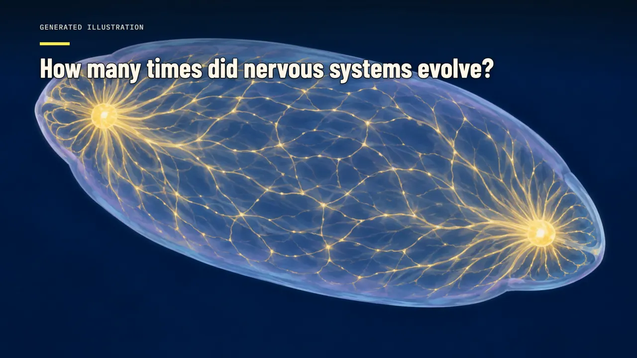
Loh's bigger idea is evolutionary: that two older nervous systems were pushed together. To judge that, it helps to know how strange early nervous systems were. Start with comb jellies. A 2023 study of gene order on chromosomes placed them as the sister group to all other animals.
In other words, their line may have branched off first, even before sponges. In 2014, the comb jelly genome led its authors to suggest that its nervous system likely has an independent origin. In 2023, scientists mapped the nerve net of a one-day-old sea walnut, a common comb jelly, with an electron microscope. Five of its nerve net neurons were fused, sharing one continuous membrane, with no gaps between them.
The authors called it a syncytial nerve net. Science News reported that comb jellies might even be a mosaic of two nervous systems of different evolutionary origins. Findings like these feed the idea that neurons may have evolved twice. Joseph Ryan of the University of Florida is skeptical.
We're dealing with close to a billion years of evolution, he said. In that much time, a single original nervous system could turn into something very strange. Leslie Babonis of Cornell says there's a lot of evidence that neurons evolved once, in the common ancestor of all animals. Max Telford of University College London says it's entirely plausible that sponges have no nervous system because they lost it.
So the question is open: two origins, or one origin with a lot of change. Jellyfish add another layer. Loh describes them as having two physically separate nervous systems at different ends of the body. The research literature describes it a bit differently.
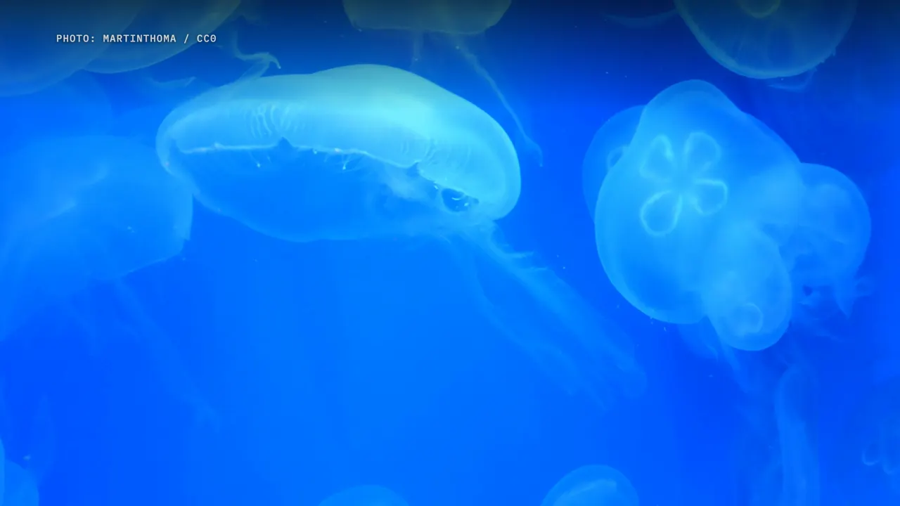
In many jellyfish, a centralized control system works alongside more diffuse nerve nets. Hydra, a tiny freshwater relative, has one nerve net in its outer layer and another in its inner layer, and they differ in anatomy and function. Either way, some animals run more than one nervous network in the same body. In 2016, Detlev Arendt's group proposed a story for how brains arose from nets like these.
An early nerve net grew two control centers, at opposite ends of the body. One, the apical nervous system, controlled general body physiology. The other, the blastoporal nervous system, coordinated feeding and movement. They proposed that these centers expanded and fused into the nerve cord and brain of animals with a left and a right side.
It sounds a lot like the Stanford story. But the 2026 study doesn't show that Arendt's two centers match its two starter populations. And on whether neurons evolved once or twice, Arendt himself told Scientific American he was undecided. A sea anemone study adds one more clue.
In the anemone Nematostella, a gene from the SIX3 family switches on at the end opposite the mouth. That's the same gene family the Stanford team used to mark the front of the brain. It's part of a long debate over which end of these animals matches our head. As a 2013 article put it: which came first, the head or the brain?
For timing, Smithsonian puts our split from jellyfish ancestors at around 600 to 700 million years ago. The split from acorn worm ancestors came roughly 550 to 600 million years ago.
Hindbrain, midbrain, forebrain: the hindbrain in segments
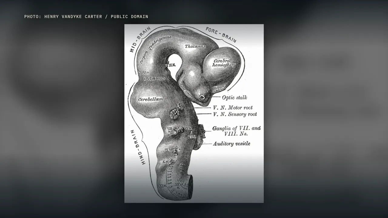
Back to the hindbrain, because the way it's built is unusual. In the embryo, the hindbrain is briefly divided into a row of segments called rhombomeres. A standard review counts seven; some schemes number them up to eight. Each segment is a lineage-restricted compartment: cells largely stay in their own segment.
It's another lineage boundary in development, though not the one the Stanford team studied. A family of genes called Hox switches on along the hindbrain in ordered, nested domains, like stair steps. The edges of those domains line up with the segment borders. And the layout of the cranial nerves, the nerves of the face, jaw and throat, can be traced back to the segments they came from.
Break one Hox gene, and the segments change. Mice lacking Hoxa1 lose rhombomere 5, and rhombomeres 4 and 6 fuse. Without Hoxb1, rhombomere 4 takes on the character of rhombomere 2. Lose a whole group of these genes, and motor neurons in rhombomeres 5 and 6 develop abnormally.
This system is old: the same Hox network for hindbrain segments is found in lampreys, near the base of the vertebrate tree. Chordates that aren't vertebrates have the nested Hox pattern but no hindbrain segments. Marianne Bronner, an author of that lamprey study, is also on the 2026 paper. And rhombomeres 5 and 6 are exactly what the Stanford team aimed for next.
Hindbrain motor neurons in a dish
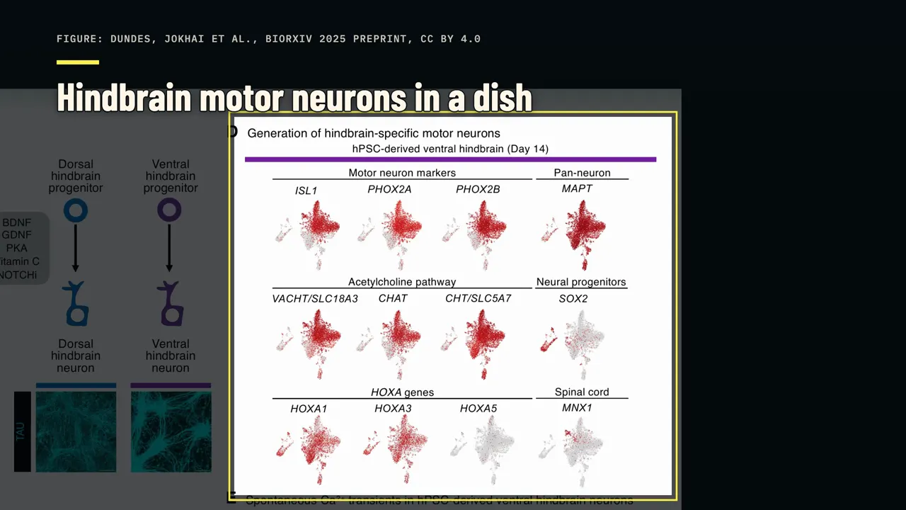
Starting from back-type cells, the team grew motor neurons with the markers of rhombomeres 5 and 6. The cells carried four hindbrain markers: HOXA1, HOXA3, PHOX2A and PHOX2B. Spinal cord markers such as HOXA5 and MNX1 were barely there. The cells also made the machinery for acetylcholine, the chemical motor neurons use to signal muscles.
They fired electrical signals called action potentials, flickered in calcium imaging, and responded to light-triggered switches. Those functional tests were small: four cells for each electrical test and ten cells for the calcium imaging, each from a single experiment. Stanford's release calls this the first time human pluripotent stem cells became functional hindbrain motor neurons in the lab. But the word first needs narrowing.
In 2018, a team in Rome made human cranial motor neurons by forcing on three genes: Ngn2, Isl1 and Phox2a. Those cells were electrically active too. The published abstract is more modest than the release: it calls these neurons hitherto difficult to generate in a dish. Nature's news story called it a difficult feat.
The Stanford route is different: it follows development, through the back-type starter cell, instead of forcing genes on. So why do hindbrain motor neurons matter? They run through the cranial nerves to the face, jaw, tongue and throat. The brainstem, also called the bulbar region, controls the muscles needed for swallowing.
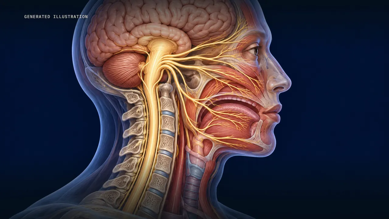
ALS attacks both the upper motor neurons in the brain and the lower motor neurons in the brainstem and spinal cord. In about a third of people with ALS, the first symptoms are in these bulbar muscles. About 75 percent of people with classic ALS eventually have some problems swallowing, speaking and chewing. Most die from respiratory failure, usually within three to five years after symptoms begin.
The most common form of SMA, spinal muscular atrophy, is caused by defects in both copies of a gene called SMN1. It damages nerve cells in the brainstem and spinal cord that control muscles for speaking, walking, breathing and swallowing. Loh's point is practical. Researchers can't sample living motor neurons from a patient's hindbrain to watch how these diseases attack them.
Growing these neurons in a dish gives researchers a way to investigate the diseases. To be clear, the paper reports no disease experiments yet. It's a new research tool, not a treatment. Funding included the NIH and the Spinal Muscular Atrophy Foundation.
The lizard brain myth, and one brain
You may have heard of the triune brain, or the lizard brain. The idea, made popular by Paul MacLean in the 1960s, is that newer brain parts were layered on top of older ones as animals evolved. A 2020 paper for psychologists answered it in its title: your brain is not an onion with a tiny reptile inside. It says the layered view has long been discredited among neurobiologists, and urges psychologists to drop it.
Its key point: all vertebrates have the same basic brain regions, forebrain, midbrain and hindbrain. So don't read the new study as an old reptile hindbrain with a new mammal forebrain on top. The split it describes may reach back past reptiles and mammals, to ancestors we share with acorn worms. It's about where cells come from, not a ladder of animals.
And again, it's front and back, not left and right. So, is your brain actually two separate organs? It's one brain, with distinct regions working together. What the experiments suggest is that this one brain is built from two beginnings.
Whether those beginnings were once two separate nervous systems is still only an idea. And whether the two starter populations are truly committed inside a living embryo is what scientists are arguing about now. One brain, two beginnings, and an open argument.
Watch on YouTube
Sources
- The study: Jokhai, Dundes et al., Nature Neuroscience 2026, doi.org/10.1038/s41593-026-02433-7
- Preprint (CC BY): Dundes, Jokhai et al., bioRxiv 2025, doi.org/10.1101/2025.07.02.662771
- Stanford Medicine news release, 18 Sep 2026 (med.stanford.edu); Stanford Report, 18 Sep 2026
- Nature news, Lynne Peeples, 18 Sep 2026, doi.org/10.1038/d41586-026-02943-1
- Ars Technica, John Timmer, "Contrary to some reports, you don't have two brains", 23 Sep 2026
- Smithsonian, Margherita Bassi, 22 Sep 2026; Nautilus, Kristen French, 18 Sep 2026; Live Science, Kenna Hughes-Castleberry, Oct 2026
- Nieuwkoop's model: Gerhart 1999, Int J Dev Biol 43:605
- Otx2 and Gbx2: Millet et al. 1999, doi.org/10.1038/43664
- Acorn worm patterning: Lowe et al. 2003, Cell
- Comb jellies: Schultz et al. 2023, doi.org/10.1038/s41586-023-05936-6; Moroz et al. 2014, Nature; Burkhardt et al. 2023, doi.org/10.1126/science.ade5645; Science News, 20 Apr 2023; Scientific American, 4 Jan 2024
- Jellyfish nerve nets: Satterlie 2011, doi.org/10.1242/jeb.043687; Jokura & Jékely 2026, EMBO J (PMC13482580)
- Arendt, Tosches & Marlow 2016, doi.org/10.1038/nrn.2015.15
- Head and brain origins: Mejia 2013, doi.org/10.1371/journal.pbio.1001484
- Hindbrain segments and Hox: Alexander, Nolte & Krumlauf 2009, Annu Rev Cell Dev Biol 25:431; Parker, Bronner & Krumlauf 2014, doi.org/10.1038/nature13723
- Cranial motor neurons by gene induction: De Santis et al. 2018, Stem Cell Research
- ALS and SMA: NINDS fact sheets (ninds.nih.gov)
- Triune brain: Cesario, Johnson & Eisthen 2020, doi.org/10.1177/0963721420917687
- Figures, images and video credits
- Study figures: Dundes, Jokhai et al., bioRxiv 2025 preprint, CC BY 4.0
- Wikimedia Commons, CC BY 4.0: Amin et al., Nesnad, Nistor et al., Stephen Thorpe, Zihao Wang
- CC BY 3.0: Ben Skála, Holger Krisp, Jack Perks
- CC BY 2.5: Giudicelli, Özbudak, Wright & Lewis
- CC BY 2.0: Smithsonian Environmental Research Center
- CC BY-SA 4.0: Brandon Antonio Segura Torres & Priscilla Vieto Bonilla, Charles J. Sharp, Eric Polk (Monterey Bay Aquarium)
- CC BY-SA 2.0 fr: Rama
- CC0: Allan Harris, Edlow et al. (Massachusetts General Hospital, 7 Tesla MRI), MartinThoma, Obsidian Soul
- Public domain: Gray's Anatomy (1918) plates by Henry Vandyke Carter; U.S. Fish and Wildlife Service
- Illustrative stock footage from Pexels (Pexels License), including laboratory, microscope, animal, and abstract scenes. This footage stands in for and does not depict the experiments or animals discussed in this video.
- Narration is synthetic. Illustrations marked "Generated illustration" on screen are AI-generated, not microscope images or records of real events. This video describes research; it is not medical advice and reports no treatment.